 KnowThis
Key Takeaways
Key Takeaways
- 1The cornea does roughly two-thirds of the eye's light-bending work, and the lens fine-tunes the rest — together they focus an image on the retina that is upside-down and reversed left-right.
- 2The retina's rod cells handle low-light and motion vision while cone cells handle color and fine detail, and it's the brain — not the eye itself — that flips the inverted image and constructs the sight you actually perceive.
- 3Common vision problems like nearsightedness and farsightedness happen because the eye focuses light slightly in front of or behind the retina rather than directly on it, which corrective lenses fix by adjusting where that focus point lands.
The concept
Understanding that the retina's raw image is upside-down — and that your brain, not your eye, flips it — is the single detail that makes the rest of how vision problems and corrective lenses work click into place.
The image projected onto your retina is upside-down and reversed left-right. Why don't you perceive the world that way?
Worked examples
Example 1: The eye's total focusing power, in diopters (baseline case)
Example 2: Nearsightedness and corrective lens power (edge case / variation)
A nearsighted (myopic) eye focuses distant light slightly in front of the retina instead of directly on it. What kind of corrective lens fixes this, and why?
Example 3: Why reading glasses become necessary with age (real-world / applied case)
The lens's ability to change shape and add extra focusing power for close-up objects — accommodation — relies on a small ring of muscle and a lens that's still flexible enough to bulge when that muscle contracts. Starting in the early-to-mid 40s for most people, the lens gradually stiffens, a normal age-related change called presbyopia, and the eye loses its ability to add that extra near-focusing power even though the muscle controlling it still works normally. This is why so many people who never needed glasses for distance vision suddenly need reading glasses, typically in the +1.50 to +2.50 diopter range, specifically for close-up tasks — it's a lens-flexibility problem, not the same kind of refractive error as childhood nearsightedness or farsightedness.
How it works (visual)
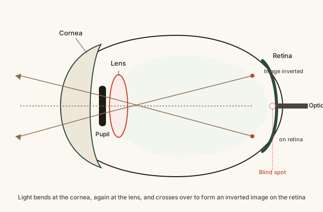
Follow a single ray of light from outside the eye: it bends sharply at the curved cornea, passes through the pupil opening, bends again at the lens for fine adjustment, and crosses over other rays before landing on the retina — that crossing-over is exactly what flips the image upside-down and left-right on the retina itself. Note where the optic nerve exits at the back of the eye: no photoreceptors exist at that exact point, which is the anatomical blind spot each eye has.
Common mistakes
Common Mistakes
Thinking the eye works like a screen that displays a finished picture.
→ The retina doesn't display anything — it's a light-detecting sensor. The actual 'picture' you consciously experience is constructed by the brain's visual cortex from the signals the retina sends.
Assuming both eyes and both types of retina cells (rods and cones) do the same job equally in all lighting.
→ Cone cells need much more light than rod cells to function, which is why color and fine detail fade first in dim light while motion and shape detection, driven mostly by rods, remain relatively strong.
Believing nearsightedness and farsightedness are the same underlying problem with different names.
→ They're opposite focusing errors — nearsightedness focuses light in front of the retina (needs a negative-diopter lens), farsightedness focuses it behind the retina (needs a positive-diopter lens).
Common misconception
“The image your eyes capture is right-side up, the same way you perceive the world.”
The optical image the eye actually projects onto the retina is inverted — upside-down and mirrored left-right — a direct, unavoidable result of how a converging lens system bends light to a focal point. The retina sends that inverted raw signal down the optic nerve exactly as captured. It's the brain's visual cortex that reprocesses and flips the signal, constructing the upright image you consciously perceive. In other words, "seeing right-side up" is a feat of neural processing, not of eye optics — the eye's optical image never stops being upside-down.
If a correctly working eye's retina always receives an upside-down, mirrored image, why doesn't the world look upside-down to you?
Try it yourself
What to do next
What to do next
- Find your own blind spot: close one eye, focus on a fixed point, and slowly move a small object off to the side at arm's length until it briefly disappears.
- Next time you're in a dim room, notice how colors look washed out while shapes and motion are still easy to detect — that's rod cells taking over from cone cells.
- If you wear glasses, check the diopter (D) number on your prescription and connect it to how strongly your lenses bend light.
- Read the related entry on Aging & the Human Lifespan to see how presbyopia fits into the body's broader pattern of age-related change.