 KnowThis
Key Takeaways
Key Takeaways
- 1The average adult has about 5 liters of blood, made up of plasma, red blood cells, white blood cells, and platelets, each with a distinct job in transport, defense, or clotting.
- 2The heart pumps blood through two connected loops — a pulmonary circuit to the lungs for oxygen, and a systemic circuit to the rest of the body — and cardiac output (heart rate x stroke volume) is the direct measure of how much blood it moves per minute.
- 3Blood is always red, at every oxygen level; veins only look blue through skin because of how light wavelengths are absorbed and scattered, not because deoxygenated blood is actually blue.
The concept
That heart rate and stroke volume relationship isn't just a definition — it's the exact number that changes when you exercise, and understanding it explains why your heart pounds so much harder during a workout than it does at rest. The nutrients that plasma carries out to the capillaries only reach the bloodstream in the first place because of how the digestive system breaks food down and absorbs it — circulation is the delivery system, not the source.
Cardiac output equals heart rate multiplied by stroke volume. During exercise, heart rate roughly doubles from 70 to 150 beats per minute. What happens to cardiac output?
Worked examples
Example 1: Resting cardiac output, with real numbers (baseline case)
Example 2: Cardiac output during intense exercise (edge case / variation)
Trained endurance athletes often have resting heart rates well below the typical 60-100 bpm range, sometimes in the 40s. What's the most likely explanation?
Example 3: Reading a blood pressure measurement (real-world / applied case)
A blood pressure reading like 120/80 mmHg reports two numbers: 120 is the systolic pressure, the peak pressure in the arteries as the heart's left ventricle contracts and pushes blood out, and 80 is the diastolic pressure, the lower pressure in the arteries as the heart relaxes and refills between beats. Both numbers are shaped by cardiac output and by how much resistance the blood vessels offer to flow — stiffer or narrower arteries raise resistance and push both numbers higher. This is why blood pressure is measured as two numbers rather than one: they capture the circulatory system under two very different conditions within the same heartbeat cycle, and consistently high readings on either number are what public health agencies track as hypertension, a major modifiable risk factor for heart disease and stroke.
How it works (visual)
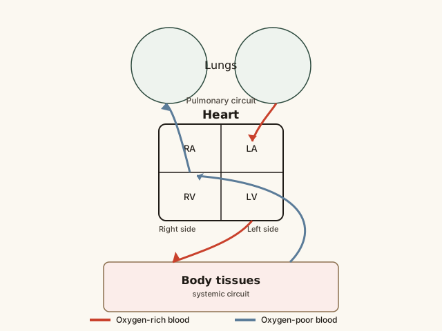
Follow the blue arrows first: oxygen-poor blood enters the heart's right side, gets pumped to the lungs, picks up oxygen, and returns to the heart's left side — that's the short pulmonary loop. Then follow the red arrows: oxygen-rich blood leaves the heart's left side, travels through the entire body delivering oxygen at the capillaries, and returns as oxygen-poor blood to the right side — that's the much longer systemic loop. Both loops run continuously and simultaneously with every single heartbeat, not one after the other.
Common mistakes
Common Mistakes
Believing deoxygenated blood is actually blue inside the body.
→ Blood is red at every oxygen level, just a darker red when oxygen-poor. Veins look blue through skin due to how skin and tissue absorb and scatter different light wavelengths, not because the blood itself changes color.
Thinking a higher heart rate alone always means more blood is being pumped.
→ Cardiac output depends on both heart rate and stroke volume together. A very fast heart rate with too little filling time between beats can actually reduce stroke volume enough to limit total output.
Assuming the two blood pressure numbers measure the same thing at different intensities.
→ Systolic (top number) and diastolic (bottom number) measure pressure during two distinct phases of the heartbeat — contraction and relaxation — not a simple 'stronger vs weaker' version of the same measurement.
Common misconception
“Blood turns blue when it runs low on oxygen, which is why veins look blue through the skin.”
Blood is red at every oxygen level because of hemoglobin, the iron-containing protein that gives red blood cells their color — it's simply a brighter red when oxygen-rich and a darker, more maroon red when oxygen-poor. It never turns blue inside the body. Veins appear blue (or greenish) through skin because of how skin and underlying fat absorb and scatter light: blue wavelengths penetrate and reflect back out of skin more effectively than red wavelengths do at the depth veins typically sit, creating a blue appearance even though the blood itself never changes to that color. If you've ever seen blood drawn during a blood test, it's visibly red the entire time, regardless of which vein it came from.
If deoxygenated blood is actually dark red rather than blue, why do veins appear blue through the skin?
Try it yourself
What to do next
What to do next
- Take your resting pulse for 60 seconds and plug it into the cardiac output calculator above using a typical 70 mL stroke volume to see your own rough resting output.
- Next time you check a blood pressure reading, connect the two numbers to the heart's contraction phase (systolic, top) and relaxation phase (diastolic, bottom) rather than treating them as one combined score.
- Notice that visible veins on your hands or wrists still look blue or greenish, and remember that's a skin light-scattering effect, not the actual color of the blood inside.
- Read the related entry on Muscles, Bones & Movement to see how the circulatory system supplies working muscle with the oxygen it needs during exertion.